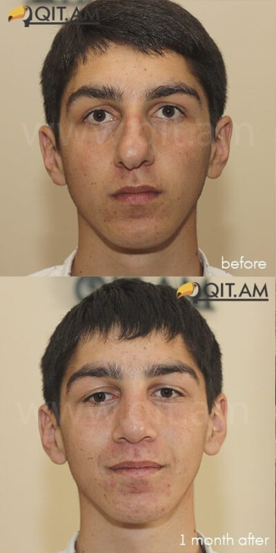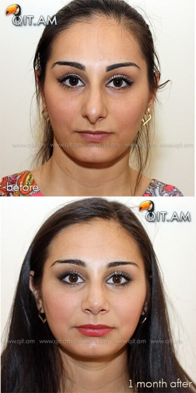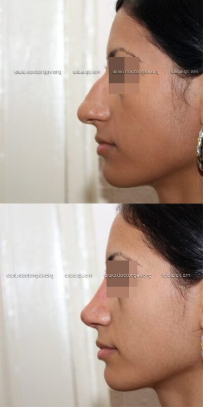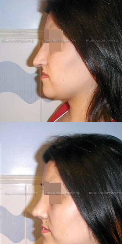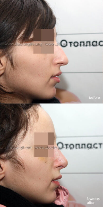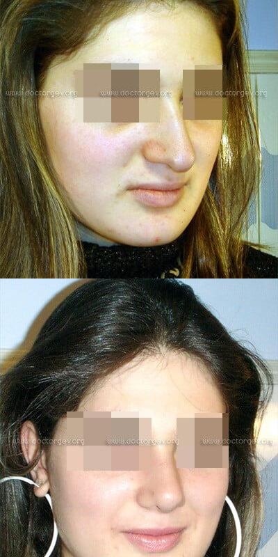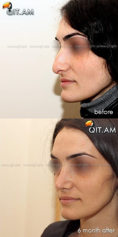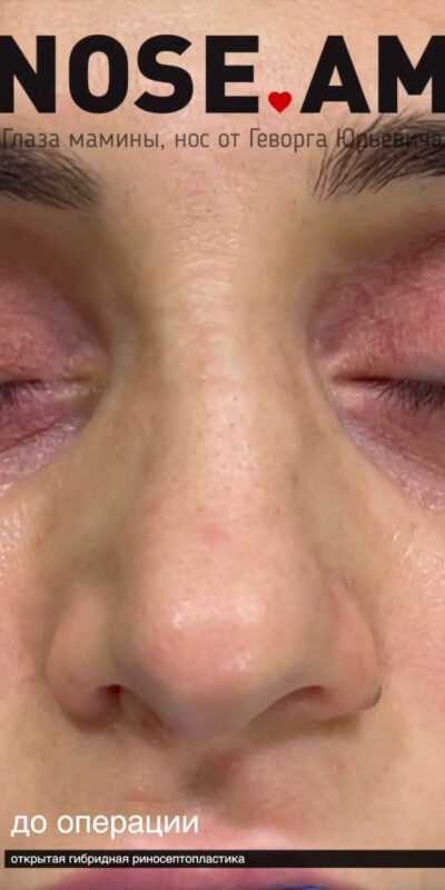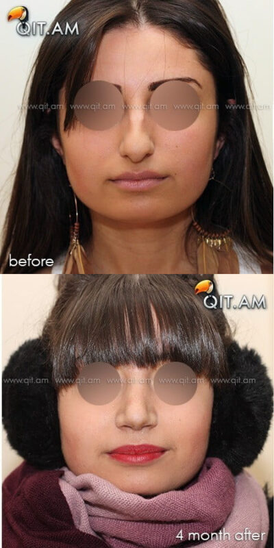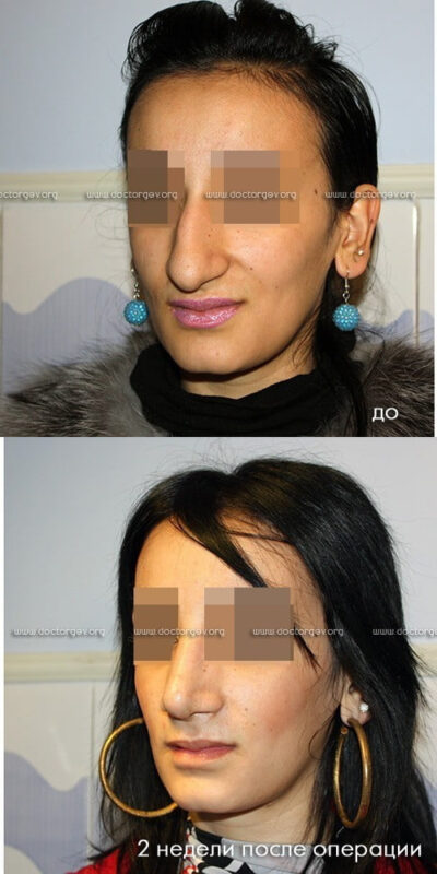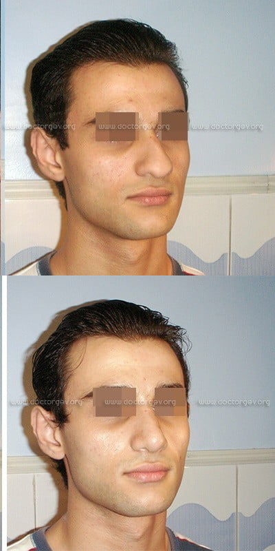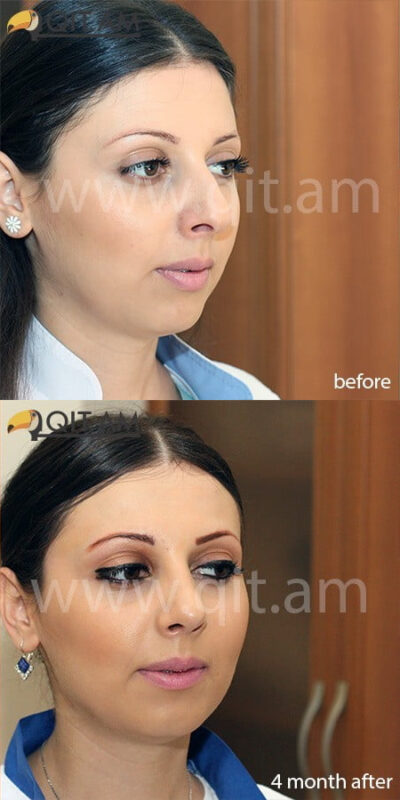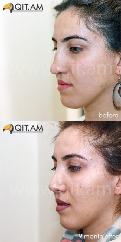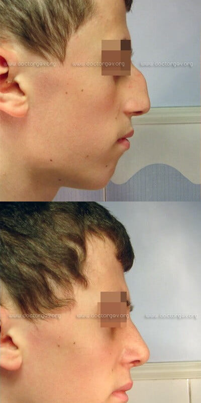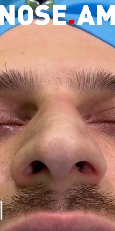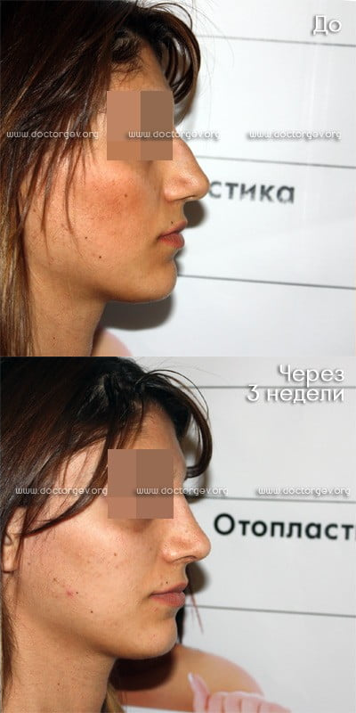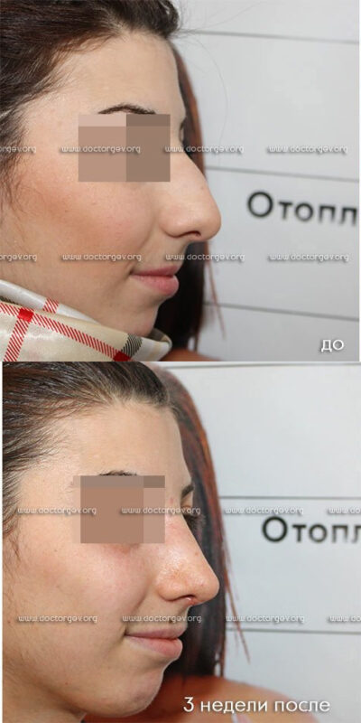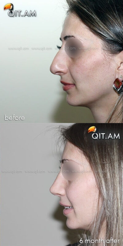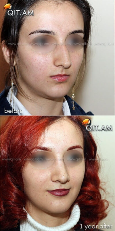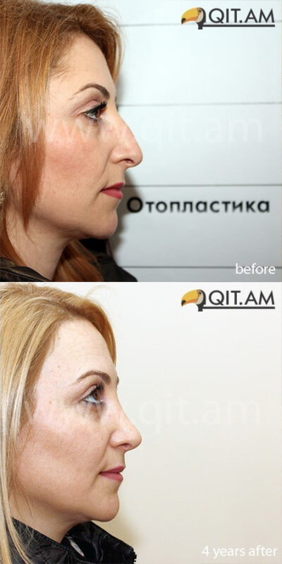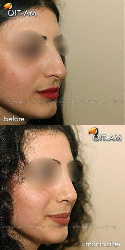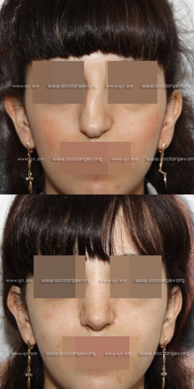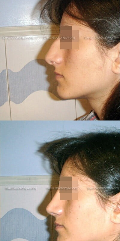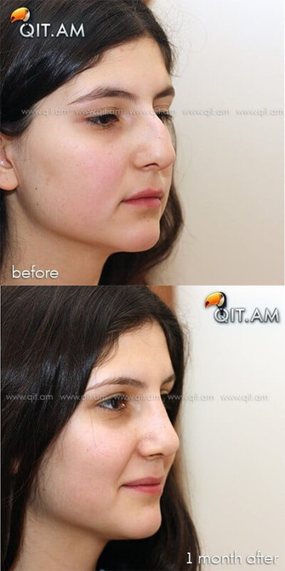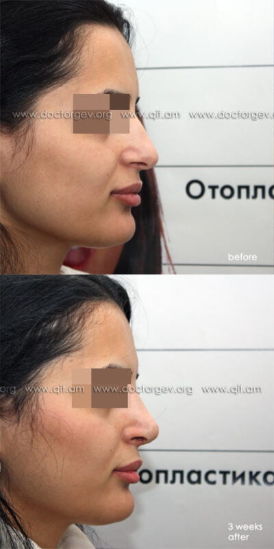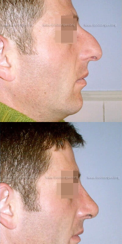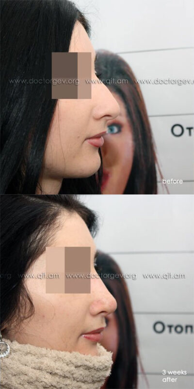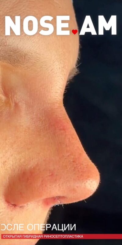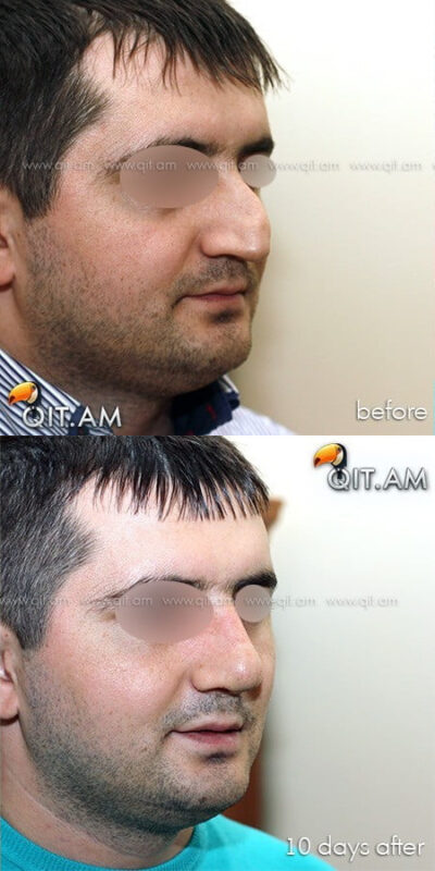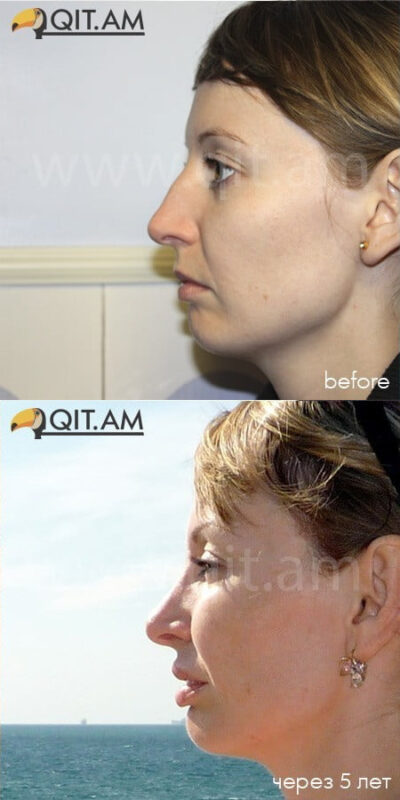

Rhinoplasty is perhaps one of the most complex facial surgical procedures. Other surgeries may be anatomically challenging, require substantial physical strength, or last until the surgeon is completely exhausted, but rhinoplasty is a surgery at the intersection of science and art. Every individual case requires a meticulous assessment of imperfections, the selection of an appropriate execution method, a thorough action plan, and a refined execution technique.
Any surgical operation can cause complications; after all, only the surgeon who does not operate makes no mistakes. Knowledge of potential complications and consequences is essential to make the right decisions, reduce the number of such cases, minimize their severity, and treat them should they occur.
Some rhinoplasty complications are related to anesthesia and are not described in this article. Anaphylactic reactions to general or local anesthesia call into question the knowledge and skills of the anesthesiologist, surgeon, and assistants.
Rhinoplasty complications can be divided into 4 categories:
- Occurring during surgery;
- Occurring immediately after surgery;
- Early;
- Delayed.
What is a complication?
In general, a complication can be described as an unexpected medical or surgical event during or after surgery that requires separate attention. Such cases are easily recognized, whereas aesthetic complications are much more difficult to identify because they are based on the surgeon’s vision and the patient’s perception of their own body. The latter factor can take various forms:
- Patient’s appearance: how do others see them? Society has certain norms of beauty and unattractiveness. Applying these standards to themselves, the patient may feel vulnerable;
- Patient’s perception of reality: how do they see themselves? Does this feeling match how others see them?
- The degree of importance the patient attaches to their appearance: some may not think about it at all, while others attach immense importance to their look;
- The degree of patient satisfaction with their appearance: dissatisfaction can range from mild discontent and concern to dysmorphic disorders leading to obsessive states. Approximately 2% of clients in plastic surgery clinics require a psychiatric evaluation.
Aesthetic perception is difficult to define, so the outcome of the surgery is always unpredictable. It depends on current fashion, media influence, cultural and ethnic characteristics.
From a practical standpoint, the beauty of the nose is based on three indicators: the ratios of width and height of frontal, transverse, and basal defects. Based on this, a three-dimensional model of the nose is created. The goal of rhinoplasty is to improve the existing harmony without disrupting the functional structure.
Complication Rates
According to data recorded in the literature, the number of complications in nasal surgery reaches 4–18.8%. For each individual surgeon, this figure decreases as experience accumulates. Complications related to the skin and soft tissues occur in 10% of cases. On average, severe systemic or life-threatening consequences arise after 1.7–5% of surgeries. Intracranial complications are extremely rare.
Description
Clinical manifestations of rhinoplasty complications can be classified as follows:
- Functional;
- Infectious;
- Aesthetic;
- Psychological;
- Specific.
Intraoperative Complications
Excessive Bleeding
This may be a consequence of congenital or acquired coagulopathy. Such conditions must be identified before surgery. If excessive bleeding occurs unexpectedly, an urgent hematologist consultation is required. As a rule, acquired coagulopathy is triggered by medication, most commonly aspirin. Its use must be suspended 2 weeks prior to surgery.
Mainly, the cause of excessive bleeding is fibrinolysis. This occurs due to abnormal activation of the fibrinolytic system, causing clotted blood to dissolve instantly. Diagnosis requires blood tests for fibrinogen and its degradation products. To stop the bleeding, blood products, norleucine, and tranexamic acid are used. However, these substances can cause deep vein thrombosis and pulmonary embolism.
Also, blood stasis during surgery can lead to excessive bleeding, complicating the surgeon’s work. This occurs in 0.3–1% of cases.
Tears of Mucochondrial Flaps
Patient and careful execution of the surgery usually prevents such complications; nevertheless, they can occur if the nose has been previously traumatized or operated on.
Unilateral tears heal on their own, but bilateral symmetrical tears can lead to septal perforation and subsequent complications. Such tears are closed immediately during surgery. Improper closure can lead to adhesion formation and nasal airway obstruction, requiring subsequent surgical intervention.
Skin Tears
This complication is possible during skin incision, especially in the area of the nasal vault. The surgeon can avoid this only by being extremely cautious. A skin tear will lead to scarring, so sutures must be placed with minimal tension. If a scar still appears, further therapy may be required.
Burns
These can occur due to equipment malfunction or surgeon error. Therefore, special care is required when using a cautery tool. If a burn occurs, various measures are taken depending on the circumstances. Skin necrosis will lead to scarring.
Disruption of the Bony Pyramid
Disruption can occur when removing a bony hump using an osteotome, especially if the patient has had prior nasal trauma or if the vomer (a flat, trapezoidal bone forming the posterior part of the nasal septum) or ethmoid bone was damaged during previously performed surgeries. In such cases, filing with a rasp is recommended.
Correction requires careful positioning of the fragments and ensuring permanent internal and external fixation during healing.
Disarticulation of the Upper Lateral Cartilage
This complication can occur when filing the bone with a rasp. Bilateral disarticulation forms an inverted-V deformity, while unilateral disarticulation leads to asymmetry in the middle third of the nose. Placing tissue grafts will eliminate ventilation symptoms and aesthetic defects.
Complications After Osteotomy
“Depression”. This deformity is a consequence of a fracture in the upper thick part of the bone at the frontonasal junction. An attempt to narrow the cut bone will lead to lateralization of the upper segment. The fracture must be displaced lower down the nasal bone.
“Open Roof”. If the lateral segments do not converge with the nasal dorsum, the resulting gap will become visible and palpable. If left unaddressed, the mucous membrane will grow into the overlying soft tissues and create pressure. Correction involves centering the nasal dorsum and aligning the lateral segments. The cause of an “open roof” can be:
- A skull bone fracture during osteotomy;
- Inability to center the fragments;
- Excessive packing in the nose.
“Step-off” Deformity. It forms during a unilateral osteotomy performed too far from the midline of the nasofacial sulcus, creating a visible hump on the side of the nose. Correction involves a repeat osteotomy.
Perinasal Trauma
During osteotomy, especially on previously traumatized noses, there is a high probability of reopening old fractures. These bones are highly susceptible to external impact. A surgeon’s error may take effect immediately or manifest as a subsequent infection. Orbital hemorrhage or panniculitis poses a threat to vision and requires immediate medical attention. Trauma to the nasolacrimal system is also possible, which may require opening a pocket in the nasal cavity. There have also been cases of infraorbital nerve damage. Complications related to skull damage are described below.
Immediate Postoperative Complications
Breathing Obstruction. Blood suctioning after extubation can cause laryngospasm. In this case, muscle relaxants, reintubation, or lung ventilation may be required. Nasal tampons or splints can also obstruct breathing. Therefore, special care must be taken when placing them.
Anaphylaxis. The probability of anaphylaxis arises when using antibiotics during surgery. Cases of anaphylactic shock from bacitracin tampons and latex are known.
Visual Impairment. After injection of local anesthetics and vasoconstrictors, temporary or permanent visual impairment is possible. The cause may be vasospasm or thromboembolism causing ocular ischemia. If symptoms persist, a doctor must be consulted.
Early Complications
Bleeding. Complications (rhinoplasty rarely causes them) occur in 2–4% of cases. Typically, the source of bleeding is localized using vasoconstrictors. Cauterization of the torn vessel is also possible. Tampons and antibiotic dressings may be required.
Septal Hematoma. Daily suctioning will be required to clear the passage of blood. Some specialists resort to incision and placement of a drainage device. To avoid an abscess in the septum, antibiotics are used.
Infections
Wound Infections. Such complications after rhinoplasty occur in only 2% of cases. Local panniculitis, abscess, or granuloma are eliminated with antibiotics or drainage. The occurrence of infection can also be triggered by the use of foreign materials.
Sepsis. It is important to realize the danger of this complication after rhinoplasty. An exacerbation of the infection can lead to acute circulatory failure and multiple organ dysfunction. Treatment options include antibiotics, hormones, and circulatory failure therapy.
Toxic Shock Syndrome. Postoperative fever, vomiting, diarrhea, low blood pressure without obvious blood loss, and an erythematous rash are signs of this syndrome. In severe cases, the condition can worsen. After a few days, desquamation of the palms and soles begins. Toxic shock syndrome toxin-1, produced by Staphylococcus aureus bacteria, is the cause of the disease. Carriers of these bacteria make up 18–50% of the healthy population.
Measures taken during treatment:
- Removal of nasal tampons, cleaning the bacterial environment;
- Antibiotic intake;
- Body examination;
- Aggressive circulatory system therapy.
Toxic shock syndrome in nasal surgery occurs in 0.016% of cases, with a mortality rate of 11%.
Subacute Bacterial Endocarditis. Every patient with heart murmurs is at risk. It is necessary to monitor oral health and take prophylactic antibiotics. If such a complication occurs, blood tests are needed.
Intracranial Infections. The proximity of the skull bone edges and traumatic manipulations during rhinoplasty increase the likelihood of meningitis, subdural empyema, and intracranial abscesses. If symptoms such as headaches, lethargy, high fever, or cranial nerve twitches appear, a doctor should be consulted.
Acute and/or Chronic Sinusitis. It can be a consequence of rhinoplasty and requires endoscopic surgical intervention.
Suture Dehiscence. This usually goes unnoticed. Adhesions may form, but they will also gradually heal. However, dehiscence of a transfixion incision requires urgent intervention, otherwise a scar forms.
Persistent Edema. The consequences of rhinoplasty can include initial swelling and periorbital bruising, which persist for up to 10 days. The severity characterizing edema after rhinoplasty depends on the complexity of the osteotomy, tools used, duration of surgery, excessive use of tampons, postoperative vomiting, or high blood pressure. To avoid all the above and prevent edema after rhinoplasty, a dressing is applied immediately after osteotomy, dexamethasone is administered intravenously during surgery, the head must remain elevated after its completion, a cold compress is applied to the nose, and blood pressure must be under constant control. Persistent edema and numbness of the nasal tip are possible with open rhinoplasty. Edema after rhinoplasty can last up to several months.
Skin Necrosis. Excessive damage and unwise use of a cautery device can lead to skin necrosis. The consequences of rhinoplasty cause impaired blood supply and infections. A dressing applied too tightly can lead to the same result. In case of necrosis, debridement and re-healing are necessary. Subsequently, to get rid of the scar, local hormonal injections, dermabrasion, laser resurfacing, and even surgical reconstruction are used.
Bone Necrosis. Necrosis of the bone or cartilage can occur, followed by infection, displacement, or associated disorders (aesthetic or functional). In such cases, the infection is first brought under control with antibiotics, and then debridement is performed. Reconstructive surgery may be required in the future.
Cardiovascular Insufficiency. In elderly patients and patients with heart disease, nasal tampons can cause hypoxia. Oxygen therapy is recommended in such cases.
Cerebrospinal Fluid (CSF) Rhinorrhea. The frequency of such complications is extremely low. Most often, rhinorrhea is triggered by prior trauma or the presence of congenital bony defects. Most leaks heal on their own. If this does not happen, intra- and extracranial surgical techniques are applied.
Contact Dermatitis. Disorders occur in patients with high sensitivity as a reaction to applied dressings. Treatment involves their removal and the administration of antihistamines or hormonal drugs.
Nasal Passage Obstruction. Edema formed after surgery can lead to permanent nasal obstruction. This condition is usually associated with vasomotor rhinopathy or allergic rhinitis. Those who are not helped by medical treatment will need surgical intervention.
Numbness and Pain. Persistent numbness and pain behind the upper incisors can be a consequence of neuropraxia of the nasopalatine nerve.
Olfactory Disturbances. Short-term impairment of smell immediately after surgery is completely justified and can have several causes, for example, edema, damage to the neuroepithelium, or the use of certain medications. Patients with prior facial trauma may be predisposed to damage to the olfactory system during osteotomy. Neurotrophic viral infections, as well as psychogenic factors, can be the reason for surgical intervention. Total loss of smell is observed in 1% of cases.
Carotid-Cavernous Fistula. This is a rare post-traumatic complication. Constant pressure fluctuation in the fistula causes eye pain, proptosis (pathological displacement of an organ or its part forward), ophthalmoplegia (paralysis of eye muscles due to damage to the oculomotor nerves), visual impairment, and murmurs. These symptoms indicate a diagnosis that can be confirmed by angiography (a method of contrast X-ray examination of blood vessels).
Reassurance Seeking. Some patients require constant reassurance from the surgeon that the nasal passage obstruction will pass, the senses of taste and smell will return, the tip of the nose will change its position, and the swelling will subside.
Early Psychological Complications. It is not uncommon for patients to experience short-term bouts of anxiety or depression, which can last up to 6 weeks.
Delayed Complications
Hypertrophic Scars. They can ruin the result of any brilliantly performed rhinoplasty. Skin loss due to infection or necrosis becomes a true disaster. It is necessary to attempt to reduce the size of the scar using hormonal injections. Next, dermabrasion, lasers, and surgical intervention are used.
“Pollybeak” Deformity. This type of deformity consists of the drooping of the nasal tip. The cause lies in improper correction of the cartilaginous dorsum or nasal septum (hard pollybeak) or in the accumulation of excess tissue during scar formation (soft pollybeak). Correction consists of reducing the cartilaginous dorsum and septum and/or excising soft tissues.
Synechia Formation. The formation of adhesions or sticking occurs when two raw surfaces come into contact. Stenting is used for prevention. Endoscopic excision is used for elimination.
Septal Perforation. Such a complication can occur in 3–24.5% of cases. For small holes, surgical closure can be used. In general, there are many execution methods and techniques. If none of them yields results, septal buttons are used.
Nasal Valve Collapse. This complication is a consequence of an overly aggressive technique of trimming the lateral cartilage. Valve collapse will cause breathing difficulties.
Nasal Passage Narrowing. This is an extremely severe complication, and it is associated with the excision of an excessive amount of tissue from the inside of the nostril. Narrowing makes breathing difficult and causes constant discomfort. Reconstructive surgery can correct the situation.
Bossae Formation. As a rule, they form on the tip of the nose in 2% of cases. While bilateral symmetrical humps can represent an aesthetically pleasing phenomenon, a bossa on one side of the nose requires surgical intervention. Therefore, during rhinoplasty, it is necessary to ensure absolute symmetry and equality of the lateral cartilage remnants. Most often, bossae are formed due to failure to maintain symmetry, the use of destructive rhinoplasty techniques, thin skin, or the formation of overly large scars.
Recurrent Meningitis. Constant bouts of meningitis can be a consequence of the failure to detect a hidden fistula. In addition to stopping the meningitis itself, its cause must be sought.
Oleogranuloma. Non-absorbable fatty materials used during surgery can trigger an inflammatory process (called oleogranuloma, paraffinoma, oil granuloma, sclerosing lipogranulomatosis). Other causes should be ruled out using computed tomography. In case of surgical intervention, recurrence is possible.
Dorsal Nasal Cyst. Nasal mucosa displaced into subcutaneous layers leads to this rare complication. Surgical intervention is possible.
Aesthetically Unsatisfactory Result
Insufficient or excessive correction of nasal imperfections leads either to the preservation of the existing defect or to the formation of a new one. In the latter case, functional disturbances may also arise. Some newly formed defects are only apparent and are corrected only after an accurate diagnosis. In general, secondary rhinoplasty should not be performed within 12 months after the first surgery.
Such defects can occur together or individually and usually affect the width, height, or depth parameters of various parts of the nose.
Upper Third Defects
- Deep Nasofrontal Angle. Various implants and grafts are used for correction.
- Shallow Nasofrontal Angle. Deepening can be done by removing the procerus muscle. If the cause lies in the bone, then osteotomy or partial removal is used.
- Widening of the Upper Third. The cause may be improper medialization of the nasal bones after osteotomy or skull bone fractures. Correction includes aligning the septum and repeat osteotomy.
- Hump of the Upper Third. Correction involves reducing the defect, usually by filing.
- Over-reduction of the Upper Third. The resulting depression needs to be corrected. If a part of the septum is missing, it must be restored.
- Asymmetry of the Upper Third. Causes can be uneven remnants of the nasal bone, asymmetrical healing, or septal deviations.
Middle Third Defects
- Widening of the Middle Third. Usually, it is a consequence of the fusion of bone and cartilage and the subsequent widening of the upper third of the nose. However, this defect may only be apparent if there is a drooping of the nasal tip.
- Hump of the Middle Third. Typically, this is a consequence of a pollybeak deformity. Correction is performed by removing excess cartilage or soft tissue.
- Depression in the Middle Third. Augmentation and, if necessary, restoration of the septum are performed.
- Asymmetry of the Middle Third. The cause may lie in unequal remnants of the bone, unilateral displacement of the cartilage, or asymmetrical healing.
Lower Third Defects
- Widening and Hump of the Lower Third. This occurs when the base of the lower part of the cartilage is destroyed.
- Wide or Boxy Nasal Tip.
- Narrow or Pinched Nasal Tip. The cause may be the destruction of the cartilage end. In addition, bossae formation or nasal valve collapse may occur. The tip of the nose is restored using graft techniques. Breathing problems are considered separately.
- Asymmetric Nasal Tip. May be the result of a breakdown in the cartilage structure. Correction depends on the cause. Graft techniques can be used.
- Overprojected Nasal Tip.
- Wide Columella. Usually, this is an initial defect not corrected during rhinoplasty. Excess soft tissues are removed from the columella.
- Hanging Columella. The cause can be deeply positioned “crura” of the septum, their removal, and subsequent scar formation. The correction method depends on the cause.
- Alar Hooding. The cause can be an excessive excision of the septum crura. Correction is performed only after an accurate diagnosis.
- Alar Rim Defects. Excessive removal of the lateral crura deprives the nostrils of support, causing them to collapse and obstruct breathing.
- Nostril Asymmetry. The cause may be the columella or the alae.
- Retracted Nasolabial Angle. The cause may be the excessive removal of the septal cartilage base. Grafts are used for correction.
- Long Nasolabial Distance. This defect may be apparent due to an elevated nasal tip. Otherwise, the removal of excess soft tissue or correction of the nasal dorsum is possible.
Other
Graft/Implant Migration. Such complications include resorption, displacement, or rejection of the implanted material. Allogeneic grafts are much more prone to rejection and infections. If antibiotics do not help, the implant is removed, after which correction is performed anew.
Disproportionate Nose. It looks unharmonious and is the consequence of a talentless surgeon’s work. After assessing the current situation, reconstructive rhinoplasty is performed.
Maxillofacial Defects. A brilliant rhinoplasty can be ruined if existing maxillofacial defects remained unnoticed before surgery. All components of both jaws, lips, and the condition of teeth must be examined in advance.
Secondary rhinoplasty is required in 5–15% of cases. Any corrective surgeries are performed with the probability of the need for further corrections.
Persistent Psychological Complications. Many studies have been conducted on this topic, the results of which are completely contradictory. Some argue that rhinoplasty patients have mental disorders and a predisposition to personality disorders. Others insist that such people have no signs of mental pathologies. Some studies even prove that after surgery people become calmer, their anxiety, hostility, and paranoid syndromes disappear, and self-esteem increases. Even patients with unstable mental health receive a positive effect from rhinoplasty. Nevertheless, in some individuals, the psychological balance can still be disrupted.
Dental Complications. During surgery, the neurovascular system of the teeth can be damaged, leading to pulp necrosis. Treatment must be carried out by a dentist.
Gustatory Rhinorrhea. Improper restoration of nerve tissues damaged during surgery leads to gustatory rhinorrhea. Treatment can be difficult; sometimes antihistamines help.
Adjuvant Disease. This is an autoimmune disease caused by hypersensitivity to the implanted material. Often, the cause can be a genetic predisposition. In some patients, symptoms can be eliminated by removing the implants. Otherwise, they consult a rheumatologist.
Lacrimal Fistula. Due to the proximity of the lacrimal drainage system to the osteotomy site, its damage is possible. However, this happens extremely rarely.
Enophthalmos and Silent Sinus Syndrome. Such complications can be a consequence of septorhinoplasty.
Patient Dissatisfaction
Aesthetic surgeries can have four outcomes:
- Satisfied patient – satisfied surgeon;
- Satisfied patient – dissatisfied surgeon;
- Dissatisfied patient – satisfied surgeon;
- Dissatisfied patient – dissatisfied surgeon.
The cause of the surgeon’s dissatisfaction is most often their own perfectionism, whereas a patient’s dissatisfaction can have many reasons, each of which is unique. The surgeon must carefully approach patient selection and examine their psychological status.
No surgical operation is immune to complications. This obliges the surgeon to be prepared for them, to know prevention measures and correction techniques. Patients must also be informed about potential complications to weigh all possible risks before surgery.
The surgeon, in turn, minimizes complications through careful patient selection (both from a medical and psychological standpoint), identifying the type of defect, and choosing the correction method.
Compliance with the doctor’s recommendations will help avoid postoperative nasal deformities and many functional disorders.
Successful rhinoplasty is the result of consolidated efforts between the doctor and the patient, requiring not only the surgeon’s precise craftsmanship and high qualifications, but also the patient’s disciplined adherence to the rules of the rehabilitation phase!
Author: Elena Ushakova, https://www.1nep.ru